Nasogastric Tube Removal Procedure⁚ A Comprehensive Guide
This comprehensive guide outlines the protocol for the nasogastric tube removal procedure. It emphasizes patient safety and comfort, providing a step-by-step approach. Proper documentation and post-removal care are also covered. This ensures timely and safe tube removal, aiding patient recovery;
Indications for NG Tube Removal
Nasogastric (NG) tube removal is indicated when the tube is no longer necessary for its original purpose. This can include situations where the patient’s gastrointestinal function has returned sufficiently to allow for oral intake, meaning they can adequately consume nutrients and fluids without assistance. Resolution of the condition that necessitated NG tube placement, such as bowel obstruction or ileus, is a primary indication.
Furthermore, if the NG tube was used for gastric decompression or drainage, its removal is appropriate once the drainage has significantly decreased or resolved altogether. In cases where medication administration was the primary reason for NG tube insertion, removal can be considered once the patient is able to take medications orally without difficulty or complications.
Clinical assessment plays a crucial role in determining readiness for NG tube removal. This assessment should include evaluating the patient’s ability to protect their airway, their level of consciousness, and their overall clinical status. The decision to remove the NG tube should be made by a qualified healthcare professional, taking into account the patient’s individual needs and circumstances. Finally, successful completion of weaning protocols, if implemented, indicates readiness for removal.
Pre-Removal Assessment and Preparation
Prior to NG tube removal, a thorough assessment is crucial. First, confirm the physician’s order for removal, ensuring it aligns with the patient’s current condition and treatment plan. Evaluate the patient’s respiratory status, including their ability to protect their airway and cough effectively. Assess bowel sounds to determine gastrointestinal motility, indicating readiness for oral intake. Check the patient’s level of consciousness and ability to follow instructions, as cooperation is essential during the procedure.

Explain the removal procedure to the patient, addressing any concerns or anxieties they may have. Provide clear and concise instructions, emphasizing the importance of relaxation and deep breathing. Gather all necessary supplies, including gloves, tissues, a clean basin, and a disposable pad to protect the patient’s clothing. Ensure proper lighting and adequate space to perform the procedure comfortably. Position the patient in a semi-Fowler’s position to facilitate breathing and prevent aspiration.
Prepare for potential complications by having suction equipment readily available in case of vomiting or respiratory distress. Assess the integrity of the NG tube, noting any signs of damage or deterioration. Consider administering oral hygiene to freshen the patient’s mouth before removal. A well-prepared environment contributes to a smooth and safe NG tube removal process.
Necessary Supplies for NG Tube Removal
Prior to commencing the NG tube removal procedure, gathering all necessary supplies is paramount for efficiency and patient safety; A clean, non-sterile pair of gloves is essential to maintain aseptic technique and protect the healthcare provider from potential exposure to bodily fluids. Tissues are needed to wipe the patient’s nose and mouth during and after the procedure, ensuring comfort and hygiene. A disposable waterproof pad should be placed over the patient’s chest to protect their clothing from any accidental spills or drainage.
A clean basin or emesis basin should be readily available to collect any gastric contents that may be expelled during the removal process. A measuring cup with water and a straw can be used to offer the patient sips of water to ease any throat discomfort experienced after removal. Assess the integrity of the NG tube, noting any signs of damage or deterioration.
Additionally, a sterile lubricant may be required to ease the removal. Suction equipment should be readily available in case of vomiting or respiratory distress. Finally, ensure proper lighting and adequate space to perform the procedure comfortably. Having all these supplies organized and within reach streamlines the procedure and minimizes any potential delays.
Step-by-Step NG Tube Removal Procedure
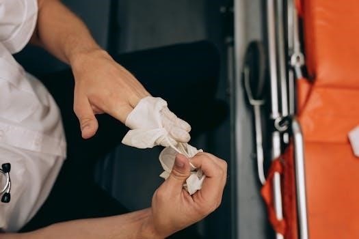
The NG tube removal process requires careful attention to detail. First, verify the physician’s order to remove the NG tube. Explain the procedure to the patient, ensuring they understand what to expect and encouraging them to relax. Position the patient upright or in a high Fowler’s position to minimize the risk of aspiration. Don non-sterile gloves and place a disposable pad on the patient’s chest.
Next, disconnect the NG tube from the suction or feeding apparatus. Gently flush the tube with a small amount of air (typically 10-20 mL) to clear it of any residual contents. Instruct the patient to take a deep breath and hold it. While the patient holds their breath, gently and steadily remove the NG tube. Wrap the removed tube in the disposable pad.
Offer the patient tissues to wipe their nose and mouth. Provide oral care, if needed. Dispose of the used supplies according to hospital policy. Document the procedure, including the patient’s tolerance and any observations. Monitor the patient for any signs of discomfort or complications following the removal. Ensure the patient can tolerate oral fluids before resuming a regular diet.
Patient Positioning During Removal
Optimal patient positioning is crucial during NG tube removal to enhance comfort and minimize potential complications. The preferred position is typically the upright or high Fowler’s position (sitting upright at a 60-90 degree angle). This position leverages gravity to reduce the risk of aspiration during and immediately after the procedure. Elevating the patient’s head and torso helps to keep the airway clear and facilitates easier breathing.
If the patient cannot tolerate a fully upright position, a semi-Fowler’s position (30-45 degree angle) may be considered. However, extra caution should be taken to monitor for any signs of respiratory distress or aspiration. In cases where the patient is unable to sit up at all due to medical conditions, the lateral decubitus position (lying on their side) might be necessary. In this case, the patient’s face should be turned towards the side, allowing easy drainage of any fluids.
Regardless of the chosen position, ensure the patient is properly supported and comfortable. Explain the importance of remaining still during the removal process to prevent injury. Proper positioning contributes significantly to a safe and smooth NG tube removal experience.
Monitoring the Patient During and After Removal
Close monitoring of the patient is paramount both during and after NG tube removal to promptly identify and manage any potential adverse reactions. During the removal process, observe the patient for signs of discomfort, gagging, coughing, or any changes in respiratory status. Assess their facial expressions and verbal cues for pain or anxiety.
Post-removal monitoring should include assessing the patient’s respiratory rate, oxygen saturation, and breath sounds. Observe for signs of aspiration, such as coughing, wheezing, or shortness of breath. Encourage the patient to take deep breaths and cough to clear any secretions. Evaluate the patient’s ability to swallow and speak without difficulty. Check for any nasal irritation, bleeding, or discomfort. Assess the patient’s level of consciousness and orientation.
Vital signs should be monitored regularly for at least the first hour after removal, and then periodically as indicated by the patient’s condition. Document all observations and interventions in the patient’s medical record. Prompt recognition and management of any complications are essential to ensure patient safety and well-being following NG tube removal.

Potential Complications of NG Tube Removal
While generally a safe procedure, NG tube removal carries potential complications that healthcare providers must be aware of. Nasal irritation, including minor bleeding or discomfort, is a common occurrence. Epistaxis, or nosebleeds, can result from trauma to the nasal mucosa during removal. Patients may also experience a sore throat or hoarseness due to irritation of the pharynx and larynx.
A more serious, though less frequent, complication is aspiration, where gastric contents enter the lungs. This can lead to pneumonia or other respiratory problems. Careful monitoring during and after removal is crucial to detect and manage aspiration promptly. Another potential issue is re-insertion difficulty if the tube needs to be replaced later. The nasal passages may become inflamed or traumatized, making subsequent insertion challenging.
Rarely, patients may experience vasovagal reactions, leading to a drop in heart rate and blood pressure. This can cause dizziness or fainting. Always have emergency equipment readily available. Proper technique and patient assessment can minimize these risks.

Post-Removal Care and Instructions
Following NG tube removal, providing appropriate post-removal care and clear instructions is essential for patient comfort and preventing complications. Immediately after removal, offer the patient oral hygiene, such as rinsing the mouth with water or using a mouthwash, to alleviate any lingering taste or discomfort. Assess the patient for any signs of nasal irritation, sore throat, or difficulty swallowing. Mild discomfort is common, but persistent or severe symptoms warrant further evaluation.
Encourage the patient to take small sips of water initially to ensure they can tolerate oral intake without difficulty. Gradually advance the diet as tolerated, starting with clear liquids and progressing to soft foods. Monitor for any signs of aspiration, such as coughing, choking, or shortness of breath. Educate the patient on these signs and instruct them to report any concerns promptly.
Advise the patient to avoid strenuous activities or excessive coughing immediately after removal, as this may irritate the nasal passages or throat. Provide written instructions regarding dietary recommendations, potential complications, and contact information for any questions or concerns. Schedule a follow-up appointment, if necessary, to assess the patient’s overall recovery and address any lingering issues.
Documentation of the NG Tube Removal Procedure
Thorough documentation of the NG tube removal procedure is a critical component of patient care and ensures continuity of information among healthcare providers. The documentation should include the date and time of the removal, as well as the indication for removal, such as resolution of the medical condition requiring the tube or patient tolerance of oral intake.
Record the patient’s tolerance of the procedure, noting any discomfort, bleeding, or other adverse reactions experienced during removal. Document the appearance of the removed NG tube, including any unusual findings such as kinks, blockages, or signs of deterioration. Note the volume and characteristics of any gastric contents aspirated prior to removal, including color, consistency, and pH level, if measured.
Include details regarding any difficulties encountered during the removal process and the interventions implemented to address them. Document the patient education provided, including instructions on oral hygiene, dietary recommendations, and potential complications to watch for. Finally, record the patient’s response to the procedure immediately following removal and any specific post-removal orders or follow-up plans. This comprehensive documentation serves as a valuable reference for future care decisions.


